English
Fukasaku Eye Institute
Welcome
Fukasaku Eye Institute welcomes you as our patients and our guests. We are honored to have repeatedly received the distinction of being sought out as the top eye surgical center by local, national, and international television, news, educational programs, print publications and celebrity patients. We will do our best for you to get your best visual acuity.
Our Mission
Our mission is to provide patients with the highest quality eye care available in the world, and to advance the field of ophthalmology through education, clinically relevant research and compassionate services.
Cataract
Cataracts are the leading cause of vision loss among adults age 55 and older.
The Fukasaku Eye Institute has compiled this information to help you better understand them.
What are cataracts?
A cataract is a cloudy area in the lens of the eye. A normal lens is clear and lets light pass to the back of the eye. A cataract blocks some of the light. As a cataract develops, it becomes harder for a person to see.
Symptoms of a cataract include: blurred driving or reading vision, sensitivity to light, glare, and halos around lights, change in glasses prescription and/or distorted images.
Types of cataracts:
A nuclear cataract occurs in the center of the lens. It may actually cause a slight improvement in near or reading vision, called “second sight.” This “second sight ” vision disappears as the cataract worsens.
A cortical cataract begins as wedge-shaped spokes in the cortex (outer portion) of the lens. When the spokes approach the center, they interfere with the transmission of light and cause glare and loss of contrast acuity.
A subcapsular cataract starts as a small opacity under the capsule (the outer membrane of the lens), and develops slowly. Vision is not significantly affected until the cataract is well developed. Diabetes, retinitis pigmentosa, and steroids are common causes of this type of cataract.
Cataract Surgery is the most commonly performed medical treatment for people 65 and older in the United States. Once a cataract reduces your vision, there are no medications, eye drops, glasses or exercises that will reverse it. Surgery, through a tiny incision, is the only way to remove a cataract. The cataract is fragmented and removed with gentle suction, and a new, permanent lens (an intraocular lens implant) is put in its place. The surgery typically uses no sutures, involves no bleeding, and is virtually painless. Although visual recovery may take up to several months to stabilize, most people can resume normal activities within a day or two.
Please contact the Fukasaku Eye Institute for an examination. Vision should not be compromised at any age.
cataract is a cloudy formation on the crystalline lens (the eyes natural lens) which may prevent a clear image from forming on the retina. Symptoms range from glare around head lights at night to needing more light when reading.
Benefits for the patient:
The procedure requires only local anesthesia (eye drops) and takes approximately 5 minutes per eye.
The intraocular lens alleviates the patient’s need for glasses both for reading and distance.
Return of visual function is rapid and most normal activities can be resumed the next day.
Patients resume their lives with a full range of vision in varying light conditions.
This procedure eliminates the need for future cataract surgery.
Refractive Lens Exchange
While LASIK corrects refractive errors of the cornea, a refractive lens exchange replaces the natural lens of your eye with an advanced multifocal intraocular lens (especially Multifocal IOL), allowing for clear vision far away, up close and in between. Additionally, patients undergoing a refractive lens exchange procedure will maintain lens clarity as they age. Any secondary opacifications can be cleared up with a simple YAG laser treatment. Refractive lens exchange works best for patients who are hyperopic (farsighted) or myopic (nearsighted), and for whom LASIK is not a viable alternative.
During the refractive lens exchange, a small incision is made at the edge of the cornea. A special instrument is inserted through the incision to gently break up and remove the natural lens. The intraocular lens implant is then inserted into place with aeronautically designed haptics (small rigid holders). The entire refractive lens exchange procedure is completed in about 10 minutes, and visual recovery is rapid.
Most patients notice significantly improved vision within 48 hours and note only mild discomfort during this initial healing time. There is no sensation of having an implant in your eye other than clear vision without glasses or contact lenses.
Fukasaku Eye Institute welcomes you as our patients and our guests. We are honored to have repeatedly received the distinction of being sought out as the top refractive center by local, national, and international television, news, educational programs, print publications and celebrity patients.
We have thus been entrusted to make it possible for you to cast aside one of life’s crutches, your eyeglasses. In providing you with this liberation, we celebrate with you your sense of elevated comfort, capability and confidence.
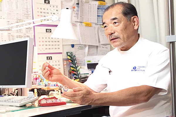
Our philosophical commitment fuels our dedication to being the best for each and every one of our trusting patients. In this regard, we are proud to provide you with our celebrated center, its staff, our standard-setting level of care and, of course, our surgeon, Dr.Hideharu Fukasaku.
Multifocal IOL
ReZoom・, Technis multi IOL/
Intra-ocular lens. Full-range vision and greater independence from glasses!
New Technology: The ReZoom・IOL and Technis multi IOL (intra-ocular lens) is a second-generation refractive multifocal acrylic IOL that provides patients with greater independence from glasses, compared to traditional (monofocal) IOL’s. It features Balanced View Optics・technology that distributes light over five optical zones for enhanced restoration of visual function, providing distance, computer and reading vision for increased independence from glasses. ReZoom・lenses are so effective, 92 percent of its recipients never or seldom need to wear glasses again.
Conditions it treats, for those who are candidates:
Nearsightedness: Also known as myopia, is the inability to see clearly at a distance. It is the result of an eyeball that is too long or the outside surface (the cornea) is too curved. Nearsightedness can be inherited or caused by the stress of too much close vision work.
Farsightedness: Also known as hyperopia, is the inability to see objects that are close up. It occurs when light entering the eye focuses behind the retina, instead of directly on it. This is caused by a cornea that is flatter, or an eye that is shorter, than a normal eye. Many children are born with farsightedness, and some outgrow it as the eyeball lengthens with normal growth.
Presbyopia: Presbyopia usually begins affecting vision at or around the age of 40. In presbyopic eyes, the natural crystalline lens of the eye loses its ability to “zoom,” meaning its ability to switch from seeing objects at a distance to seeing objects up close. A result of being presbyopic is having to hold things farther away to read or a dependence on reading glasses or bifocals.
Cataracts:
A cataract is a cloudy formation on the crystalline lens (the eyes natural lens) which may prevent a clear image from forming on the retina. Symptoms range from glare around head lights at night to needing more light when reading.
Benefits for the patient:
The procedure requires only local anesthesia (eye drops) and takes approximately 10 minutes per eye.
The intraocular lens alleviates the patient’s need for glasses both for reading and distance.
Return of visual function is rapid and most normal activities can be resumed the next day.
Patients resume their lives with a full range of vision in varying light conditions
This procedure eliminates the need for future cataract surgery.
Cornea, External Disease, and Cataract Service
The Cornea, External Disease, and Cataract Service offers consultation in the medical and surgical management of all disorders of the cornea, conjunctiva and anterior segment. We offer expertise in corneal transplantation, including high risk keratoplasty and keratoplasty in association with iris and lens abnormalities, as well as diagnosis and management of corneal ulcers, scleritis, ocular allergy and pemphigoid.
Special services include:
excimer laser therapeutic and refractive keratectomy (LASIK, PRK and PTK)
computerized video corneal topography
anterior segment photography and angiography
endothelial specular microscopy
therapeutic and refractive contact lens fittings
microbiology evaluations
immunopathology and histopathology of conjunctival and corneal disease including pemphigoid and tumors
Glaucoma service
The Glaucoma and Cataract Service provides complete diagnostic and therapeutic care for the glaucoma referral patient.
Facilities for diagnostic testing include:
automated static and kinetic Perimetry (Humphrey, FDT)
blue on yellow Perimetry (SWAP)
manual Perimetry with Goldmann and tangent screen
pachymetry
stereo disc photography
nerve fiber layer photography
Optical Coherence Tomography (OCT)
contrast sensitivity
electrophysiologic testing (multifocal ERG & VEP)
ultrasound biomicroscopy of the anterior segment
Medical and surgical therapies include:
conventional filtration surgery including trabeculectomy with or without antimetabolites (Mitomycin C)
argon, diode, and Nd:YAG lasers for cyclophotocoagulation, trabeculoplasty, selective laser trabeculoplasty, iridectomy and other laser procedures.
glaucoma implant surgery, including implants
Vitreous & Retinal Detachment
Most of the serious retinal problems which require surgery are caused by problems with the vitreous, the clear jelly-like substance which fills the space in the eye.
Posterior Vitreous Detachment
With age, the vitreous becomes more fluid, and less jelly-like. As the eyeball moves, small pockets of liquid vitreous can move around inside the vitreous cavity. This movement causes the vitreous to pull on the retina, and, with time, the vitreous can separate from the retina. This is called posterior vitreous detatchment (PVD), because it usually happens at the back (posterior) of the eye. PVD happens in most people eventually, and is rarely a problem.
Flashes and Floaters
When PVD occurs, the detached vitreous can tug on the retina. The brain interprets these tugs as flashes or large spots in the vision. The vitreous can also become stringy, and form visible strands which appear in the field of vision as floating threads or small spots and circles. A patient with these floaters should be examined, to check for other retinal damage. If there are no problems, the patient can fairly easily learn to ignore the floaters.
Retinal Tear and Vitreous Hemorrhage
Where the vitreous is securely attached to the retina, vitreous detachment may cause the retina to tear. If the retina tears across a blood vessel, there will be bleeding into the vitreous – this is called vitreous hemorrhage. Small amounts of bleeding cloud the vision, leading to the sensation of walking through a swarm of insects – more severe bleeding leads to a mass of red or black lines, and vision may become very dark. A retinal tear is a serious problem; vitreous hemorrhage is even more serious.
Retinal tears may be sealed with lasers or cryotherapy, or both, to prevent retinal detachment. Both these treatments seal the retina to the wall of the eye, repairing the tear and preventing detachment.
Retinal Detachment
A retinal tear is considered so serious because the vitreous liquid may leak through the tear, and collect under the retina. Gradually, the build up of liquid separates the retina from the wall of the eye, a condition called retinal detachment. The two major classical treatments for retinal detachment are scleral buckling – where a sponge or length of silicon plastic is placed on the outside of the eye and sewn in place, pushing the sclera toward the tear in the retina – and pneumatic retinopexy, a less severe treatment where the surgeon injects a gas bubble inside the vitreous cavity. The bubble pushes the retina against the wall of the eye, allowing the tear to seal against the eye wall. However,the recent modern vitreous surgery (vitrectomy) can be applied for every kind of retinal detachment.
Vitreous Surgery
If the retinal detachment is too severe for scleral buckling or pneumatic retinopexy, surgery to reattach the retina may be necessary. Under local anesthetic, the surgeon removes the vitreous entirely, replacing it with air or a fluid compatible with the eye. Over time, the fluid (or air) is absorbed, and replace with the eye’s own fluid. Lack of vitreous does not affect the patient’s vision.
Results of the retinal detachment surgery
In the Fukasaku Eye Institute, we have recovered 100% of the retinal detachment cases, if the eye is not underwent any surgery at another clinic. So, please consult the Fukasaku Eye Institute as soon as possible, if you feel some symptom.
New technique for minimally invasive vitrectomy
Since the technique of retinal-vitreous space surgery (vitrectomy) was first developed in the 1970’s to treat the diverse spectrum of retinal disorders, a uniform standard has existed for the instrumentarium: All instruments, such as suction and cutting tools, scissors, forceps, and laser probes, have had a uniform diameter of 20 ga (0.9 mm).
The conventional vitrectomy procedure requires that an opening is first cut in the superficial conjunctiva for introduction of instruments into the eye. Three to 4 incisions are then made in the sclera for accesses to the vitreous space. At conclusion of the procedure, sutures must be used to close the incisions in the sclera and conjunctiva. Although they dissolve within 2 to 4 weeks, the sutures can cause discomfort (feeling of a foreign body, tearing eye) and almost always produce a marked reduction in visual acuity due to warping of the cornea (astigmatism).
In 2002 the American retinal surgeon Eugene de Juan developed a procedure for minimally invasive vitrectomy. Using microcannulas and a 25 ga (0.5 mm) instrumentarium, the incisions made in the conjunctiva and sclera are so small that the wounds self-seal without sutures. This so-called 25 ga vitrectomy, however, has a number of drawbacks. Not all of the instruments can be manufactured with such a small (25 ga) diameter, while those currently available are too flexible and relatively unstable. Moreover, the incisions do not always close tightly on the first day after surgery, with the result that for several days the intraocular pressure can remain so low that sutures do after all become necessary.
The procedure of minimally invasive ‘sutureless’ vitrectomy was further refined in 2004 at the Eye Clinic of the Municipal Clinics Frankfurt-Hoechst . A novel tunnel incision technique and newly developed microcannula system enable the use of larger, more stable 23 ga (0.65 mm) instruments.
Almost all of the 23 ga instruments required for this procedure are currently available. Our experience shows that the wound closure is clearly superior to that of 25 ga vitrectomy. Since Spring of 2004, therefore, all minimally invasive retinal surgeries at our clinic in the Fukasaku Eye Institute are performed using only the 23 ga technique.
Many different types of vitrectomy can be performed with the new minimally invasive 23 ga technique. Compared to the conventional 20 ga procedure, it offers the advantages of less trauma to tissues, smaller incisions, more rapid healing, less risk of postoperative inflammatory reactions (redness, swelling), little or no discomfort, and quicker attainment of the operative goal of improved visual acuity.
In the Fukasaku Eye Institute, we perform this 23G vitrectomy technique in almost every cases of the vitreo-retinal surgery and obtain the early visual rehabilitation.
LASIK Laser Refractive Surgery and Phakic IOL
At our laser vision correction offices in Yokohama, Japan, we are committed to offering patients customized vision correction and personalized care. We firmly believe that strong doctor-patient communication leads to optimal surgical results. Therefore, our practice encourages patients to ask questions at every step of the refractive surgery process, from initial consultation to follow up appointments. We invite you to browse this website to get better acquainted with LASIK eye surgery, PRK, CK, and other treatments we offer. For more information on a specific procedure, you can attend one of our free laser eye surgery seminars at the Fukasaku Eye Institute.
Custom LASIK
.
Custom LASIK is a new and exciting advancement in the field of refractive surgery. When performing custom LASIK laser eye surgery, our Boston – based practice uses cutting edge wavefront technology to create a visual map of each of your eyes. Using this map, we are able to treat even the slightest visual imperfections, called higher-order aberrations. Custom LASIK results in a superb quality of vision, reducing minor visual imperfections such as glare, halos, and poor night vision.
If you are interested in receiving vision correction that is tailored to your individual needs, you should consider undergoing custom wavefront LASIK surgery at our Boston practice. Wavefront LASIK goes one step beyond traditional LASIK, providing treatment based on three-dimensional measurements of your eyes. In order to collect these measurements, our practice utilizes the highly advanced VISX S4 IR (Iris Registration sytem) Custom Vue system. This system analyzes your eye and creates a wavefront map that is used to guide your custom LASIK treatment.
Phakic IOL; Artiflex IOL implant surgery
LASIK is not good choice for very high myopia, especially it will lose a night good vision after LASIK. In -8 diopter or more, we perfopm phakic IOL, Artiflex IOL, implant surgery to correct very high myopia. The result is very good and patients satisfaction is very high.
Vision Rehabilitation Service
The Vision Rehabilitation Service at the Fukasaku Eye Institute helps patients with low vision or partial sight to make full use of their remaining vision at the highest level possible.
Many eye diseases and injuries, as well as birth defects, may cause low vision. The most common cause is Age Related Macular Degeneration (ARMD). Since ARMD results in only central retinal visual loss, leaving peripheral (side) vision unaffected, it is often possible to help these patients make better use of the remaining side vision. Conversly patinets with peripheral visual field loss due to hemianopia or RP can benefit from special devices developed by Dr. Peli for treating these conditions.
The Vision Rehabilitation Service provides a complete low vision evaluation that determines visual function under various conditions, and assesses the patient’s work or home visual requirements.
The service fits patients, based on the evaluation results, with one or more visual aids to help him or her perform various daily activities. The service also provides instruction and in-office training in the use of a full array of optical and non-optical aids.
The service also offers:
? a complete selection of advanced electronic visual aids (television and scanning devices);
? a Pediatric Vision Rehabilitation Program (in collaboration with our Pediatric Ophthalmology Service) including specialized low vision evaluation and rehabilitation for children and adolescents;
? a complete consulting service regarding driving rehabilitation or driving cessations, as needed.
Visual aids cannot cure or reverse partial sight. Visual aids prescribed and dispensed at the service are usually aimed at providing magnification. With proper magnification, many patients can perform tasks that were impossible without magnification. Other devices serve to expand the peripheral field.
Reading & Other Near Vision Aids
The magnification for reading may be provided by various types of spectacles, ranging from fairly common looking reading glasses to extremely strong magnification glasses. Magnification for reading and other near tasks may be provided with a variety of hand-held magnifiers. The type and strength of the magnifier prescribed are fitted individually based on the type and severity of the visual loss and the task to be accomplished. Attention is given to the patient’s limitation of manual control, dexterity or posture, which may affect the utility of the aids.
Electronic aids for magnification include various types of closed circuit TV magnifiers, ranging from the large, desktop models to small, portable (battery operated) systems. These devices provide larger magnification (30-60 times) and therefore afford the visually impaired person flexibility and comfort in reading.


